Category: infection

  • Dengue Fever in Thailand 2026: A Doctor’s Guide to Symptoms, Prevention, and Treatment

    What is Dengue Fever?

    Dengue fever is a mosquito-borne viral infection caused by four closely related dengue virus serotypes (DENV-1 through DENV-4) of the genus Flavivirus. It is transmitted primarily by the day-biting Aedes aegypti mosquito, with Aedes albopictus serving as a secondary vector. Dengue is now the most common vector-borne viral infection in tropical and subtropical regions worldwide, with over 100 countries affected across Southeast Asia, the Western Pacific, the Americas, and Africa.

    Epidemiology in Thailand

    Thailand remains one of the countries most heavily affected by dengue fever in Southeast Asia. As of March 2023, dengue is endemic in more than 100 countries, and the Americas, Southeast Asia, and the Western Pacific regions are the most seriously affected. Thailand reports tens of thousands of dengue cases annually, with significant seasonal variation. The rainy season from May through October creates ideal breeding conditions for Aedes mosquitoes, leading to dramatic surges in cases during these months.

    In Pattaya and surrounding Chonburi Province, dengue activity has historically been elevated due to the warm tropical climate, urban density, and tourism-related population movement. Dengue fever acquired in Southeast Asia typically presents with more severe hemorrhagic symptoms compared to infections acquired elsewhere, and cases can progress to dengue hemorrhagic fever or dengue shock syndrome.

    Symptoms: What to Watch For

    The incubation period after a dengue mosquito bite is typically 4 to 7 days, with symptoms lasting 2 to 7 days. Most infected individuals are asymptomatic or have only mild symptoms, but about 20% develop noticeable illness. The classic presentation includes:

    • Sudden high fever: Typically 40°C (104°F), often lasting 2 to 7 days

    • Severe headache: Especially pain behind the eyes (retro-orbital pain), which is characteristic of dengue

    • Severe muscle and joint pain: So severe that dengue was historically called “breakbone fever”

    • Nausea and vomiting: Common during the febrile phase

    • Rash: A macular or maculopapular rash that typically appears a few days after fever onset, involving the trunk and spreading outward

    • Mild bleeding: Nosebleeds, gum bleeding, or easy bruising

    • Fatigue and malaise: Profound tiredness that can persist for weeks

    Most patients recover fully within a week or so, though convalescence can be prolonged, with fatigue and weakness lasting several weeks.

    Dengue Hemorrhagic Fever and Warning Signs

    Secondary infection with a different dengue serotype carries a significantly higher risk of severe disease due to antibody-dependent enhancement. The World Health Organization now uses an updated classification system that identifies patients with dengue who have warning signs requiring careful observation:

    • Abdominal pain or tenderness

    • Persistent vomiting

    • Clinical fluid accumulation (abdominal distension, difficulty breathing)

    • Mucosal bleeding

    • Lethargy or restlessness

    • Liver enlargement greater than 2 centimeters below the right costal margin

    • Laboratory findings: increase in hematocrit greater than 20% of baseline with a rapid decrease in platelet count

    Without prompt fluid resuscitation, patients who progress to dengue shock syndrome can die within 12 to 24 hours. However, with appropriate hospital care, mortality is less than 1%.

    Diagnosis

    Early diagnosis (days 1 to 5 of illness) relies on:

    • NS1 antigen detection: Rapid antigen-based ELISA tests that can identify dengue virus non-structural protein 1 during the acute phase

    • RT-PCR: Reverse transcription polymerase chain reaction, the gold standard for detecting dengue viral RNA during the first week of illness

    After day 5, diagnosis shifts to serology:

    • Dengue IgM ELISA: Becomes positive after 4 to 5 days of illness

    • Dengue IgG ELISA: Indicates past infection or secondary infection

    Common laboratory findings include leukopenia (low white blood cell count), thrombocytopenia (platelet count below 100,000), and modest elevations of serum AST (aspartate aminotransferase) without significant liver synthetic dysfunction.

    Treatment

    There is no specific antiviral treatment for dengue. Management is entirely supportive:

    • Adequate hydration: Oral or intravenous fluids are the cornerstone of dengue management. Patients should drink plenty of fluids to maintain hydration and prevent shock from plasma leakage.

    • Paracetamol (acetaminophen): The recommended antipyretic for fever and pain relief. NSAIDs (ibuprofen, aspirin, diclofenac) should be avoided until dengue has been ruled out, as they increase the risk of bleeding.

    • Close monitoring: Patients with warning signs should be hospitalized for serial hematocrit measurements, platelet counts, and clinical observation.

    • Blood transfusion: May be needed in severe dengue with significant bleeding or profound thrombocytopenia.

    Prevention

    There is currently no universally available dengue vaccine, although several vaccines are in use in various countries. The most effective prevention strategies focus on mosquito control and personal protection:

    • Eliminate breeding sites: Empty, cover, or treat any container that collects standing water around the home — flower pots, buckets, discarded tires, water storage containers, and roof gutters

    • Use mosquito repellent: DEET-containing repellents applied to exposed skin

    • Wear protective clothing: Long sleeves and long pants, especially during daytime when Aedes mosquitoes are most active

    • Install window screens: Use screens on doors and windows, or sleep under insecticide-treated bed nets

    • Community action: Support local mosquito control programs and fogging operations

    Dr. Kijakarn notes that dengue is particularly dangerous in Pattaya due to its high tourism volume. “Many tourists are not aware of dengue risk and do not take prevention measures seriously. We see adults and children alike presenting with severe dengue during the rainy season. Anyone visiting or living in tropical Thailand should treat dengue prevention with the same seriousness as malaria prevention.”

    This article is for general information only and does not replace professional medical advice. If you are experiencing symptoms, please consult a healthcare provider.
  • Leptospirosis in Tropical Thailand: The Hidden Danger After Floods

    What is Leptospirosis?

    Leptospirosis is a zoonotic bacterial disease caused by spirochetes of the genus Leptospira. It is considered the most common worldwide infectious zoonosis, with cases reported across all continents except Antarctica. The disease affects both animals and humans, with rodents, cattle, dogs, and other mammals serving as reservoir hosts. The bacteria are shed in the urine of infected animals and can survive in warm, moist environments for weeks to months, making water and soil contaminated with infected animal urine the primary routes of transmission to humans.

    How People Get Infected

    Transmission occurs when bacteria in contaminated water or soil enter the body through cuts or scratches in the skin, through mucous membranes of the eyes, nose, or mouth, or, rarely, through ingestion of contaminated food or water. Activities that place people at high risk include:

    • Agricultural work, especially rice farming and sugarcane harvesting

    • Wading or swimming in contaminated freshwater (rivers, lakes, ponds, floodwater)

    • Occupational exposure in veterinarians, farmers, slaughterhouse workers, and sewage workers

    • Recreational activities like whitewater rafting, kayaking, and triathlons in tropical regions

    • Contact with animals or animal urine in endemic areas

    In Thailand, leptospirosis is a significant public health concern, particularly in rural agricultural communities during the rainy season. The northern and northeastern regions of Thailand report the highest incidence rates, with cases closely correlated with flooding and heavy rainfall.

    Symptoms: From Mild Flu-like to Life-Threatening

    Leptospirosis is known as “the great imitator” because its symptoms mimic many other diseases. The incubation period ranges from 2 to 30 days, typically 7 to 14 days.

    Classic (anicteric) leptospirosis — accounting for about 90% of cases — presents as a sudden-onset flu-like illness:

    • Sudden high fever: Often reaching 39°C to 40°C (102°F to 104°F)

    • Chills and rigors: Often sudden and dramatic

    • Severe muscle aches: Particularly affecting the calves, lower back, and thighs — calf tenderness is a classic finding

    • Severe headache: Often frontal and frontal-temporal

    • Nausea, vomiting, and diarrhea: Gastrointestinal symptoms are common

    • Rash: Macular or petechial, appearing in some cases

    • Conjunctival suffusion: Red eyes without purulent discharge — this is a highly characteristic finding of leptospirosis

    • Jaundice: Yellowing of the skin and eyes (absent in anicteric form)

    Severe leptospirosis (Weil’s disease) occurs in approximately 5% to 10% of cases and can be fatal:

    • Severe jaundice with liver failure

    • Acute kidney injury requiring dialysis

    • Pulmonary hemorrhage — diffuse alveolar hemorrhage can be rapidly fatal

    • Meningitis and meningoencephalitis

    • Cardiac arrhythmias and myocarditis

    • Disseminated intravascular coagulation (DIC)

    • Multiorgan failure and death (mortality in Weil’s disease is 5% to 15%, and up to 50% in severe pulmonary hemorrhagic syndrome)

    Diagnosis

    Diagnosis is challenging because early symptoms overlap with many tropical diseases. Key diagnostic tools include:

    • Culture: The definitive diagnostic method, but Leptospira grows slowly (up to 13 weeks) and requires specialized media, making it impractical for acute management

    • PCR: Detects Leptospira DNA in blood (first week) and urine (second week onward); provides rapid results

    • Microscopic Agglutination Test (MAT): The gold standard serological test, detecting agglutinating antibodies; requires paired sera showing a four-fold rise in antibody titer

    • Rapid diagnostic tests: IgM-based ELISA and lateral flow assays provide faster results but may have variable sensitivity

    • Complete blood count: May show thrombocytopenia and elevated C-reactive protein

    • Chemistry panel: Elevated creatinine (kidney injury), bilirubin, and liver enzymes

    Leptospirosis must be differentiated from malaria, typhoid fever, dengue fever, scrub typhus, viral hepatitis, and other febrile illnesses — all of which are common in tropical Thailand.

    Treatment

    • Mild disease: Oral doxycycline (100 mg twice daily for 7 days) or oral amoxicillin/ampicillin. Most patients can be treated at home with antibiotics, fluids, and supportive care.

    • Severe disease: Intravenous ceftriaxone (1-2 g daily) or IV penicillin G. Hospitalization is essential for close monitoring of renal function, fluid balance, and respiratory status.

    • Supportive care: Aggressive fluid and electrolyte management, renal replacement therapy (dialysis) if needed, and careful monitoring for pulmonary hemorrhage.

    Early antibiotic treatment — even within the first five days of illness — shortens the clinical course, reduces complications, and decreases the duration of bacteremia and bacterial shedding.

    Prevention

    • Avoid contact with potentially contaminated water: Especially during and after flooding. Do not swim or wade in floodwater, rivers, or ponds in endemic areas.

    • Wear protective clothing: Waders, boots, and gloves when working in waterlogged soil or with animals

    • Cover wounds: Use waterproof dressings on any cuts or abrasions before contact with potentially contaminated water

    • Antibiotic prophylaxis: Doxycycline 200 mg weekly during high-risk activities (e.g., rafting, military exercises in endemic areas)

    • Control rodents: Reduce rodent populations around homes and workplaces

    • Vaccination: Vaccines are available in some countries for high-risk occupational groups, but no vaccine is widely available globally

    Dr. Kijakarn emphasizes the connection between flooding and leptospirosis in Thailand. “Every major flood in Thailand is followed by a spike in leptospirosis cases. We see farmers and flood cleanup workers presenting with high fever, muscle pain, and jaundice. Anyone who has been wading in floodwater and develops fever should seek medical care immediately and specifically mention their water exposure — this dramatically speeds up diagnosis and treatment.”

    This article is for general information only and does not replace professional medical advice. If you are experiencing symptoms, please consult a healthcare provider.

  • Scrub Typhus in Southeast Asia: What Every Doctor and Traveler Needs to Know

    What is Scrub Typhus?

    Scrub typhus is an acute febrile illness caused by the bacterium Orientia tsutsugamushi, a member of the rickettsial group of pathogens. It is transmitted to humans through the bite of infected chigger mites (larval trombiculid mites) that feed on rodents, the natural reservoir of the organism. Scrub typhus is found across a vast “tsutsugamushi triangle” stretching from northern Japan and eastern Russia in the north, through southern China and the Indian subcontinent, to northeastern Australia and the western Pacific islands. It is particularly endemic in Southeast Asia, including Thailand, where it is a recognized cause of acute undifferentiated febrile illness, especially in rural and agricultural areas.

    Epidemiology in Thailand

    Scrub typhus is a significant cause of acute undifferentiated fever in Thailand, particularly in the northern and northeastern regions where rice paddies, forests, and brushy areas provide ideal habitat for the chigger mites. Cases peak during the cool, dry season from November to April, when agricultural activities such as rice harvesting bring people into close contact with chigger-infested areas.

    The disease is increasingly recognized as one of the most common causes of acute encephalitis and febrile illness with multiorgan involvement in Southeast Asia. In Thailand, scrub typhus is often underdiagnosed because its symptoms overlap with dengue, leptospirosis, malaria, and typhoid fever — all of which are prevalent in the same geographic areas.

    Symptoms: The Classic Triad and Beyond

    The incubation period is typically 1 to 3 weeks after a chigger bite. The disease has an abrupt onset with the following characteristic features:

    • Sudden high fever: Often reaching 39°C to 40°C (102°F to 104°F), with an acute, dramatic onset

    • Severe headache: Often frontal, persistent, and refractory to analgesics

    • Myalgias: Muscle aches, particularly of the back and shoulders

    • The eschar: At the site of the chigger bite, a papule develops that becomes necrotic and forms a characteristic black crusty lesion called an eschar. The groin and abdomen are the most common sites, followed by the chest and axillae. The eschar is typically painless with an erythematous halo and may have lymphangitis radiating from it. While this finding is highly suggestive of scrub typhus, it was only described in approximately 19% of a South Korean study series, and it is often located in hidden areas (groin, axillae, scalp) and can be missed on examination.

    • Rash: A maculopapular rash that typically appears around days 5 to 7 of illness, beginning on the trunk and spreading to the extremities. The rash may be subtle or absent in some cases.

    • Lymphadenopathy: Regional lymph nodes near the eschar are commonly enlarged and tender

    • Splenomegaly: Enlargement of the spleen

    Severe scrub typhus can involve multiple organ systems and may be fatal without treatment:

    • Encephalitis: Confusion, altered mental status, seizures

    • Pneumonitis: Respiratory distress, acute respiratory distress syndrome (ARDS)

    • Myocarditis: Cardiac inflammation, arrhythmias

    • Meningitis: Nuchal rigidity, photophobia

    • Hepatitis: Elevated liver enzymes, jaundice

    • Acute renal failure

    Without treatment, fever typically subsides spontaneously after about 2 weeks, but the mortality rate may be 10% to 30%. With appropriate antibiotic treatment, mortality drops below 1%.

    Diagnosis

    Diagnosis is primarily clinical, supported by laboratory and serological findings:

    • Serological assays: Indirect fluorescent antibody (IFA), enzyme-linked immunosorbent assay (ELISA), and rapid diagnostic tests using lateral flow chromatography are the mainstays of laboratory diagnosis. IFA is considered the gold standard but results take days to weeks.

    • PCR: Polymerase chain reaction amplification of Orientia genes from eschar tissue is highly effective for early diagnosis. Blood PCR is less sensitive but useful in eschar-negative cases.

    • Blood tests: Typical findings include elevated C-reactive protein, mild transaminitis (elevated AST/ALT), thrombocytopenia, and hyponatremia. Lymphocytosis and an elevated white blood cell count may be present.

    • Chest X-ray: May show interstitial infiltrates in cases with pneumonitis

    Scrub typhus must be differentiated from dengue, leptospirosis, typhoid fever, malaria, rickettsialpox, murine typhus, and other causes of acute undifferentiated fever. The presence of an eschar, lymphocytosis, and elevated C-reactive protein may help distinguish scrub typhus from dengue.

    Treatment

    Scrub typhus responds dramatically to antibiotic therapy, and treatment should never be delayed pending laboratory confirmation when clinical suspicion is high:

    • First-line treatment: Doxycycline 100 mg orally twice daily. Treatment should continue until the patient has been afebrile for at least 3 days (typically 7 to 14 days total). A rapid defervescence — often within 24 to 48 hours — is characteristic of successful treatment and serves as a useful diagnostic clue.

    • Alternative for mild disease: Azithromycin 500 mg once daily for 3 days. Shorter courses are associated with higher relapse rates.

    • Severe disease: Minocycline 100 mg intravenously twice daily, or doxycycline if IV is not available. Chloramphenicol (500 mg four times daily) is an alternative but carries a risk of relapse and serious adverse effects including aplastic anemia.

    Relapse occurs in up to 20% of cases, especially with short-course therapy or in patients who stop antibiotics too soon after fever resolves. Patients should be instructed to complete the full course of treatment and return if fever recurs.

    Prevention

    • Avoid chigger habitats: Chiggers thrive in grassy, brushy, and wooded areas. Avoid sitting on the ground in tall grass, brush, or forest floor, especially during the cool, dry season.

    • Use insect repellent: DEET-based or permethrin-treated clothing provides effective protection against chigger bites

    • Wear protective clothing: Long sleeves, long pants tucked into socks, and closed shoes when working or hiking in endemic areas

    • Shower immediately: Showering and changing clothes after potential exposure can wash off unengaged chiggers before they bite

    • Clear vegetation: Keep grass and brush trimmed around homes, especially in rural areas

    • Chemoprophylaxis: Daily doxycycline 100 mg has been used for military personnel and others with prolonged high-risk exposure, but is not generally recommended for tourists or the general public

    Dr. Kijakarn notes that scrub typhus is frequently misdiagnosed as dengue in Thailand. “Both diseases cause high fever in the same geographic areas, but the treatments are completely different. If a patient has been hiking, farming, or spending time in grassy areas and presents with fever, we specifically look for the eschar. Missing the diagnosis and delaying doxycycline can be fatal, especially in children and elderly patients. We always ask about outdoor exposure before assuming dengue.”

    This article is for general information only and does not replace professional medical advice. If you are experiencing symptoms, please consult a healthcare provider.

  • Influenza Outbreak 2026: What You Need to Know About the H3N2 Subclade K Wave

    It is mid-2026, and influenza is back with a vengeance. What started as a mild seasonal uptick has now escalated into one of the most significant flu outbreaks the world has seen in over a decade. Hospitals across Asia, Europe, and North America are reporting surges in respiratory cases that far exceed normal seasonal patterns, and public health officials are sounding the alarm.

    The current outbreak is driven primarily by a mutated strain of influenza A(H3N2), specifically a new subclade designated as K. This variant has proven remarkably effective at evading the immune protection conferred by last season’s flu vaccine. According to the U.S. Centers for Disease Control and Prevention, the 2025–2026 flu season has already caused approximately 25 million illnesses and 20,000 deaths in the United States alone — including the tragic loss of 79 children. Similar trends are being reported across Southeast Asia, where Thailand, Vietnam, and Malaysia have all reported elevated influenza activity well above the seasonal baseline.

    Dr. Kijakarn, a physician at a major hospital in Pattaya, notes that the clinical presentation of this year’s flu has some unexpected features. “We’re seeing patients who are much sicker than typical seasonal flu,” he explains. “The fever tends to run higher, the cough is more persistent, and recovery is taking longer. Many patients report lingering fatigue for two to three weeks after the acute phase has passed.”

    Why is the 2026 flu season so severe?

    Several factors are converging to make this flu season particularly challenging:

    Vaccine mismatch: The H3N2 subclade K mutation has reduced the effectiveness of the current flu vaccine. While the vaccine still provides meaningful protection against severe disease and hospitalization, its ability to prevent infection altogether has declined significantly. Experts estimate protection against infection is now around 30–40%, down from the usual 50–60%.

    Post-pandemic immune debt: After years of reduced influenza circulation during the COVID-19 pandemic, population-wide immunity to flu viruses waned. As normal travel and social interactions resumed in 2024–2025, the virus found a much larger pool of susceptible hosts than usual.

    Co-circulation with other viruses: This season has also seen unusual co-circulation of influenza with respiratory syncytial virus (RSV), adenovirus, and SARS-CoV-2. This “twindemic” scenario strains healthcare systems and makes differential diagnosis more complex.

    Delayed peak timing: Historically, flu peaks between November and January in the Northern Hemisphere. This year, the peak has been delayed into spring, keeping the virus circulating for a longer period and extending the window of transmission.

    Symptoms: How to tell flu from a cold or COVID-19

    The symptoms of the 2026 flu strain overlap significantly with common cold, COVID-19, and other respiratory infections. Here is what to watch for:

    • Sudden onset: Flu hits fast. A cold usually develops gradually over a day or two. Flu, by contrast, can leave you feeling fine one hour and debilitated the next.

    • High fever: Most adults with flu get a fever of 38°C (100.4°F) or higher. Younger children and older adults may not always develop a fever, which can delay diagnosis.

    • Body aches: The deep muscle and joint pain associated with flu is often more severe than with a cold. Many patients describe feeling like they “have been hit by a truck.”

    • Fatigue: Flu-related fatigue is profound and can last for weeks. It is distinct from the milder tiredness of a common cold.

    • Respiratory symptoms: Dry cough, sore throat, and nasal congestion are common with flu, but the cough tends to be more persistent and can last four to six weeks.

    • Gastrointestinal symptoms: While more common in children, nausea, vomiting, and diarrhea are sometimes seen with influenza, particularly with H1N1 strains.

    What should you do if you suspect you have flu?

    Get tested. Rapid influenza diagnostic tests can provide results in 15–30 minutes at most clinics. Knowing whether you have influenza, COVID-19, or another respiratory virus guides treatment decisions and helps prevent spread.

    Start antiviral medication early. Oseltamivir (Tamiflu) and baloxavir (Xofluza) are most effective when started within 48 hours of symptom onset. For patients at high risk — those over 65, pregnant women, people with chronic conditions, and young children — early antiviral treatment can prevent serious complications including pneumonia, sepsis, and death.

    Rest and hydrate. These old-fashioned measures are not optional. Your immune system needs energy and fluids to fight the virus. Pushing through flu “like a man” only prolongs recovery.

    Isolate. Influenza is highly contagious. Stay home for at least 24 hours after your fever breaks without using fever-reducing medication. In this outbreak, consider extending isolation to five days or until symptoms improve, whichever comes first.

    Prevention in the 2026 outbreak

    Even with a partially mismatched vaccine, flu shots remain the single most effective prevention tool. The WHO has recommended the same three-strain formulation for the 2026–2027 season — H1N1, H3N2, and B/Victoria lineage — which is very similar to last year’s vaccine.

    Beyond vaccination, standard preventive measures matter more than ever in this outbreak:

    • Wear a well-fitting mask (N95/KN95 or surgical) in crowded indoor spaces

    • Wash hands frequently and avoid touching your face

    • Improve ventilation in indoor spaces — open windows, use HEPA filters

    • Stay home when sick

    • Keep distance from people who are coughing or sneezing

    Dr. Kijakarn emphasizes that Pattaya, with its high volume of international visitors, is at particular risk. “Tourists who are asymptomatic or mildly ill can spread the virus to vulnerable local populations — elderly residents, young children, and immunocompromised patients. We urge visitors to get vaccinated before travel and to seek medical care promptly if they develop symptoms.”

    The 2026 influenza outbreak is a stark reminder that respiratory viruses do not disappear — they evolve. By staying informed, getting vaccinated, and practicing good hygiene, you can reduce your risk of infection and help protect the broader community.

    This article is for general information only and does not replace professional medical advice. If you are experiencing symptoms, please consult a healthcare provider.

  • Acute Febrile Illness (AFI) Causes

    Acute Febrile Illness (AFI): Causes

    Acute Febrile Illness (AFI) is characterized by a sudden onset of fever and associated systemic symptoms lasting for a short duration, typically less than two weeks. It is a common presentation in clinical settings and can be caused by a wide range of infectious and non-infectious etiologies. Proper identification of the underlying cause is crucial for effective management and treatment. This article provides a comprehensive list of all possible causes of AFI, including rare causes.

    Causes of Acute Febrile Illness (AFI)

    Table 1: Causes of AFI (Including Rare Causes)

    TypeCauseExamples
    Infectious CausesBacterial InfectionsTyphoid Fever, Pneumonia, Meningitis, UTIs, Endocarditis, Brucellosis, Tularemia, Anthrax, Q Fever, Melioidosis, Leprosy, Lyme Disease, Rickettsial infections (Rocky Mountain Spotted Fever, Scrub Typhus)
    Viral InfectionsDengue, Influenza, COVID-19, Chikungunya, Measles, Mumps, Rabies, Hantavirus, Viral Hemorrhagic Fevers (Ebola, Marburg), West Nile Virus, Zika, Crimean-Congo Hemorrhagic Fever (CCHF), Lassa Fever, Nipah Virus
    Parasitic InfectionsMalaria, Leptospirosis, Trypanosomiasis, Toxoplasmosis, Babesiosis, Schistosomiasis, Amoebiasis, Fascioliasis, Filariasis, Echinococcosis
    Fungal InfectionsCandidiasis, Histoplasmosis, Coccidioidomycosis, Cryptococcosis, Blastomycosis, Paracoccidioidomycosis, Sporotrichosis, Aspergillosis
    Non-Infectious CausesAutoimmune DisordersSystemic Lupus Erythematosus (SLE), Rheumatic Fever, Still’s Disease, Sarcoidosis, Vasculitis, Adult-onset Still’s Disease (AOSD), Polyarteritis Nodosa
    Malignancies (Neoplastic)Lymphoma, Leukemia, Renal Cell Carcinoma, Hepatocellular Carcinoma, Multiple Myeloma, Metastatic Cancers, Paraneoplastic Syndromes
    Drug-Induced FeversAntibiotics, Anticonvulsants, Chemotherapy, NSAIDs, Antiarrhythmics, Vaccines, Biologics, Illicit Drug Reactions
    Environmental CausesHeat StrokeHeat Stroke, Hyperthermia due to extreme environments, Neuroleptic Malignant Syndrome (NMS)
    Hyperthermia from ToxinsCocaine, Amphetamines, Anticholinergics, Salicylate Toxicity, Serotonin Syndrome
    Unknown CausesFever of Unknown Origin (FUO)Idiopathic Fevers, Familial Mediterranean Fever (FMF), Adult-onset Immunodeficiency Syndrome (AOIS), Periodic Fever Syndromes

    Pathophysiology

    1. Infectious Causes (Most Common)

    • Bacterial Infections:
      • Pathophysiology: Activation of immune response via endotoxins or exotoxins leading to cytokine release (IL-1, TNF-α).
      • Examples:
        • Typhoid Fever (Salmonella typhi)
        • Pneumonia (Streptococcus pneumoniae, Haemophilus influenzae)
        • Meningitis (Neisseria meningitidis, Streptococcus pneumoniae)
        • Urinary Tract Infections (E. coli)
    • Viral Infections:
      • Pathophysiology: Viral replication causing cell lysis, immune activation, and cytokine storm.
      • Examples:
        • Dengue Fever
        • Influenza
        • COVID-19 (SARS-CoV-2)
        • Chikungunya
        • Measles, Mumps
    • Parasitic Infections:
      • Pathophysiology: Immune response to parasitic antigens; inflammation and tissue damage.
      • Examples:
        • Malaria (Plasmodium spp.)
        • Leptospirosis (Leptospira spp.)
        • Trypanosomiasis (African Sleeping Sickness)
    • Fungal Infections:
      • Pathophysiology: Invasive fungal infections trigger inflammatory responses, especially in immunocompromised individuals.
      • Examples:
        • Candidiasis
        • Histoplasmosis

    2. Non-Infectious Causes

    • Autoimmune Disorders:
      • Pathophysiology: Autoimmune activation causing systemic inflammation and fever.
      • Examples:
        • Systemic Lupus Erythematosus (SLE)
        • Rheumatic Fever
        • Still’s Disease
    • Malignancies (Neoplastic):
      • Pathophysiology: Release of pyrogens from cancer cells or tumor necrosis.
      • Examples:
        • Lymphoma
        • Leukemia
        • Renal Cell Carcinoma (Paraneoplastic syndromes)
    • Drug-Induced Fevers:
      • Pathophysiology: Hypersensitivity reactions or alteration in thermoregulation.
      • Examples:
        • Antibiotics (e.g., Beta-lactams)
        • Anticonvulsants (e.g., Phenytoin)
        • Chemotherapy agents

    3. Environmental Causes

    • Heat Stroke:
      • Pathophysiology: Failure of thermoregulation leading to hyperthermia and systemic inflammatory response.
    • Hyperthermia from Toxins:
      • Pathophysiology: Direct effect on hypothalamic regulation or increased metabolic rate.
      • Examples:
        • Cocaine, Amphetamines
        • Anticholinergic overdose

    4. Unknown/Idiopathic Causes

    • Fever of Unknown Origin (FUO):
      • Pathophysiology: Persistent fever (>3 weeks) without clear etiology despite thorough investigation.

    References

    1. Mandell, Douglas, and Bennett’s Principles and Practice of Infectious Diseases, 9th Edition.
    2. Harrison’s Principles of Internal Medicine, 21st Edition.
    3. Kumar & Clark’s Clinical Medicine, 10th Edition.
    4. Fauci AS, et al. (2020). Harrison’s Principles of Internal Medicine. McGraw-Hill.
    5. Shapiro DS, et al. (2019). Infectious Disease: A Clinical Short Course. McGraw-Hill.
    6. Longo DL, et al. (2019). Fever of Unknown Origin. New England Journal of Medicine.
  • Disseminated Gonorrhea

    Disseminated Gonorrhea

    Disseminated Gonorrhea or Neisseria Gonorrhoeae. Disseminated gonococcal infection (DGI)

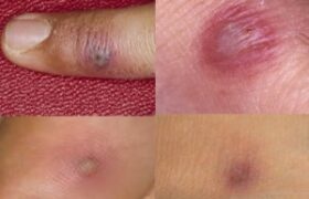
    Many of my patients with Gonorrhea( pus per urethra) present with rash , some have joint pain (Gonococcal arthritis)

    (DGI) follows untreated mucosal infection in about 0.5-3 per- cent of patients. Skin lesions are the most common manifestation of DGI and occur in 50-70 percent of patients. The eruption typically appears during the first day of symptoms and may recur with each bout of fever. The skin lesions associated with DGI begin as tiny red papules or petechiae 1-5 mm in diameter, many of which evolve rapidly through vesicular or pustular stages to develop a gray necrotic center, often on a hemorrhagic base. Papules, bullae, pustules, and hemorrhagic lesions may all be present simultaneously. The lesions tend to be scanty but widely distributed. The distal portions of the extremities are most commonly involved, with sparing of the scalp, face, trunk, and oral mucous membranes. Histologic examination will reveal local vasculitis, fibrin deposition, necro- sis, and neutrophil infiltration. Gram-stained smears of material from skin lesions infrequently reveal organisms, although most smears are positive for gonococci when examined by immunofluorescence techniques. Circulating immune complexes may play a role in the pathogenesis.